Anzeige


Wissenschaftliches Programm
|
Ab 08:00
|
Registrierung |
|
|
R
|
||
|
08:45 – 09:15
|
Gemeinsame Eröffnung |
|
|
Theatersaal
GE
|
Maurizio Calcagni, Zürich; Patricia Kammermann, Bern
|
|
Some anecdotes and insights on cartoons from a hand-drawing animation filmmaker
Dustin Rees, Zürich
|
|
|
|
09:30 – 10:30
|
Freie Mitteilungen I |
|
|
Ballsaal
SGHR-FM1
|
Astrid Schmid, Thun; Barbara Roland, Miège
|
|
|
FM60
Ist propriozeptive Rehabilitation bei Handgelenkssteifheit nach der Operation hilfreich?
Charlotte Soete, Neuchâtel
DetailsDistalradiusfrakturen (DRF) sind bei Erwachsenen häufig und machen etwa 17,5% aller Frakturen aus1. Die Rehabilitation stellt den Therapeuten vor die Herausforderung, einerseits die Schmerzen zu reduzieren und andererseits die Beweglichkeit, Kraft und Funktion zu verbessern. Komplikationen kommen dabei oft vor und werden in 27% der Fälle von Ärzten gemeldet (basierend auf Diagnosen) und in 21% von Patienten (basierend auf Symptomatik) 2. Bei DRF, die mit Osteosynthese behandelt wurden, sind Gelenksteife und an verschiedenen Stellen des Handgelenks lokalisierte Schmerzen auch nach drei Monaten postoperativ nicht ungewöhnlich. Mehrere Literaturrecherchen zur Behandlung dieser komplexen Fälle nach der Operation ergaben keine Informationen über die Behandlung. In einigen Artikeln und Büchern wird die Rehabilitation der bewussten und unbewussten Propriozeption für die Rehabilitation nach DRF diskutiert. Es handelt sich hierbei um ein weniger bekanntes Vorgehen, das für Handtherapeuten nicht einfach umzusetzen ist3. Könnte das von Mesplié4 übernommene Hagert-Programm helfen, bei einem steifen Handgelenk mehr als drei Monate nach der Operation den Bewegungsradius und die Funktionalität zu verbessern? Die Anwendung dieses Programms in zwei klinischen Fällen soll hier vorgestellt und die Implikationen für die Praxis diskutiert werden. 1Nellans, K. W., Kowalski, E., & Chung, K. C. (2012). The Epidemiology of Distal Radius Fractures. Hand Clinics, 28(2), 113‑125. https://doi.org/10.1016/j.hcl.2012.02.001 2McKay, S. D., MacDermid, J. C., Roth, J. H., & Richards, R. S. (2001). Assessment of complications of distal radius fractures and development of a complication checklist. The Journal of Hand Surgery, 26(5), 916‑922. https://doi.org/10.1053/jhsu.2001.26662 3Hagert, E. (2010). Proprioception of the Wrist Joint : A Review of Current Concepts and Possible Implications on the Rehabilitation of the Wrist. Journal of Hand Therapy, 23(1), 2‑17. https://doi.org/10.1016/j.jht.2009.09.008 4Grégory Mesplié, Josette Mesplié - Thérapie de la main : Anatomie fonctionnelle et thérapie des pathologies du poignet. (s. d.). Consulté 30 avril 2023, à l’adresse https://www.sauramps.com/product/49160/gregory-mesplie-josette-mesplie-therapie-de-la-main-anatomie-fonctionnelle-et-therapie-des-pathologies-du-poignet |
|
|
|
FM61
Motorische Kontrolle und motorisches Lernen in der Handtherapie
Vera Beckmann-Fries, Zürich; Céline Schneider, Zürich
DetailsHintergrund: In der Handtherapie werden die Prinzipien der motorischen Kontrolle und des motorischen Lernens intuitiv angewendet. Es ist jedoch sinnvoll, diese bewusst zu reflektieren und in die Therapie einzubeziehen. Nach einer Verletzung oder bei degenerativen Veränderungen müssen Bewegungen neu wahrgenommen, erlernt oder adaptiert werden, um letztendlich die volle Funktion der oberen Extremität: das Greifen, Manipulieren und Stützen, wieder durchführen zu können. «Motorische Kontrolle bezieht sich auf den Prozess, mit dem das zentrale Nervensystem Bewegungen des Körpers plant, koordiniert und ausführt.»1) Die motorische Kontrolle umfasst die Steuerung der Muskulatur und die Kontrolle von Gelenksbewegungen, um koordinierte und funktionelle Bewegungen zu ermöglichen. Sensorische Inputs und Wahrnehmung helfen, eine Bewegung auszuwählen und zu steuern. Die motorische Kontrolle spielt eine wesentliche Rolle in alltäglichen Aktivitäten, wie Gehen, Greifen von Gegenständen, Sportausübung und anderen Bewegungsabläufen. «Die Grundprinzipien des Motorischen Lernens können auf verschiedene Weise formuliert werden, aber im Allgemeinen beziehen sie sich auf die grundlegenden Konzepte, die das Lernen von motorischen Fähigkeiten steuern und beeinflussen.»1) Wiederholungen und Übungen sind Schlüsselkomponenten, wenn es darum geht, Bewegungen (neu) zu erlernen. Feedback spielt hierbei eine wichtige Rolle, da es hilft, die Leistung zu bewerten und Anpassungen vorzunehmen. Fehler machen gehört dazu, und Korrekturen können umgesetzt werden. Zudem ist es hilfreich, Bewegungen und Aktivitäten in unterschiedlichen Umgebungen auszuführen. Dies führt zur Kontextualisierung, also der Art und Weise, wie eine Bewegung in welcher Umgebung und welchem Ziel ausgeführt wird. Ziel/Implikation: Die Grundlagen der motorischen Kontrolle und des motorischen Lernens werden vorgestellt und anhand praktischer Beispiele aus der Praxis erläutert. 1)OpenAI’s ChatGPT AI language model, persönliche Kommunikation, 18.05.2023 |
|
|
|
FM62
Behandlung von Allodynien der oberen Extremität durch die Anwendung von Pyonex-Nadeln (PN)
Céline Thuler, Neuchâtel
DetailsEinleitung: Bei der Rehabilitation der oberen Extremität begegnen wir häufig Patienten, die aufgrund von Nervenverletzungen an mechanischer Allodynie leiden. Die Anwendung von PN in der Peripherie des allodynischen Bereichs erschien uns als Behandlungsansatz, den es näher zu beleuchten gilt. Es handelt sich um eine einfache, kostengünstige und für den Patienten wenig belastende Behandlung. Ziel der Studie ist es, die Wirkung der oberflächliche Afferenzstimulation (Superficial Dry Needling) bei der Behandlung von Allodynien im Bereich der oberen Extremität unter Verwendung von NP als therapeutisches Mittel zu untersuchen. Methode: Die Einzelfallanalyse wurde unter Anwendung der Single-Case-Experimental-Design-Methode durchgeführt. Wiederholte Messungen wurden im Doppelblindverfahren über 12 Wochen während 3 "ABA"-Phasen vorgenommen, wobei die Baseline (A) 3 Wochen und die Behandlung (B) 6 Wochen dauerten. Die Einschlusskriterien waren, dass der Patient seit mindestens 6 Monaten an Allodynie in der oberen Extremität leidet und dem Begutachter sowie dem Therapeuten vor Beginn der Studie unbekannt ist. Als primäre Messgrösse wählten wir die Allodynographie zusammen mit der Regenbogen-Schmerzskala und als sekundäre Messgrösse die Schmerzeinschätzung. Ergebnis: Die Ergebnisse belegen, dass die Behandlung mit PN die Grösse des allodynischen Bereichs in diesem einzelnen Studienfall verringert. Der allodynische Bereich war zu Beginn der Studie 296cm2 und nach 12 Wochen 170cm2 gross, was einer Abnahme um 43% entspricht. Der Bereich der Regenbogen-Schmerzskala betrug 169cm2 und ging auf 57cm2 zurück, hat also um 66% abgenommen. Wir beobachten auch eine Stabilisierung der Allodynographie zum Zeitpunkt der Beendigung der Nadelbehandlung, sodass wir zum Schluss kommen, dass die Anwendung der Nadeln tatsächlich eine Wirkung hatte. Die Entwicklung der Schmerzen ist weniger eindeutig, es ist keine Korrelation zwischen der Schmerzintensität, der Aufnahme der Behandlung und deren Beendigung zu beobachten. Schlussfolgerung: Diese Studie zeigt ein potenzielles Forschungsfeld auf und validiert einen möglichen neuen therapeutischen Ansatz zur Behandlung von statischen mechanischen Allodynien. Durch Studien in grösserem Massstab könnte die Beweiswirkung noch gesteigert werden. Wir könnten uns auch andere Anwendungsgebiete vorstellen, z.B. in Bereichen, die sich im proximalen Teil des kutanen Verteilungsgebiets des geschädigten Nervenastes befinden. |
|
|
|
FM63
Kinesiophobie, zu beachten in der Handtherapie? Eine Annäherung
Nadine Schulz, Zürich; Vera Beckmann-Fries, Zürich
DetailsHINTERGRUND: Die Entstehung der Kinesiophobie, also der Angst vor körperlicher Bewegung, ist komplex und tritt bei einer Vielzahl von Personen mit unterschiedlichen muskuloskelettalen Erkrankungen oder einer Vorgeschichte von Operationen auf (Huang et al., 2022). In Ihrer Arbeit zeigten Pagels et al. (2022) auf, dass Kinesiophobie bei Schulterbeschwerden beispielsweise einen negativen Einfluss auf den Therapieerfolg hat. Ähnliche Ergebnisse wurden auch von Tuna & Oksay (2018) erzielt, die Patienten nach einer Sehnenoperation an der Hand untersuchten und feststellten, dass Patienten mit stärkerer Kinesiophobie schlechtere funktionelle Resultate erzielten als Patienten mit geringerer Kinesiophobie. Trotz dieser Erkenntnisse finden sich nur wenige Studien, die sich mit der Kinesiophobie im Zusammenhang mit Handtherapie, Handverletzungen und -erkrankungen befassen. ZIEL/IMPLIKATION: Das Ziel dieser Literaturarbeit besteht darin, einen aktuellen Überblick über das Thema Kinesiophobie in der Handtherapie zu geben, offene Fragen zu identifizieren und zu untersuchen, ob es spezifische praxisbezogene Ansätze gibt. Darüber hinaus soll aufgezeigt werden, wie Kinesiophobie erfasst und gemessen werden kann und ob vorhandene Befundinstrumente auch für die Handtherapie reliabel und valabel sind. METHODE: Literaturrecherche in Datenbanken wie PubMed, Cinahl, MedLine, sowie Fachbücher RESULTATE: Resultate und daraus resultierende Erkenntnisse werden am Kongress vorgestellt. Huang, J., Xu, Y., Xuan, R., Baker, J. S., & Gu, Y. (2022). A Mixed Comparison of Interventions for Kinesiophobia in Individuals With Musculoskeletal Pain: Systematic Review and Network Meta-Analysis. Front Psychol, 13, 886015. https://doi.org/10.3389/fpsyg.2022.886015 Pagels, L., Lüdtke, K., & Schäfer, A. (2022). Kinesiophobie bei Schulterbeschwerden. Der Schmerz. https://doi.org/10.1007/s00482-022-00678-2 Tuna, Z., & Oskay, D. (2018). Fear of movement and its effects on hand function after tendon repair. Hand Surg Rehabil. https://doi.org/10.1016/j.hansur.2018.05.004 |
|
|
|
09:30 – 10:30
|
Hauptsession IMinimal Invasive hand surgery |
|
|
Theatersaal
HS1
|
Dominique Merky, Bern; Sebastian Günkel, Solothurn
|
|
Cutting-Edge Advances in Interventional Wrist Arthroscopy
Lorenzo Merlini, Paris (FR)
|
|
|
Le traitement endoscopique du syndrome du canal carpien
Maurizio Calcagni, Zürich
|
|
|
Endoskopische Nervenchirurgie – nicht nur im Sulcus
Damian Sutter, Bern
|
|
|
Minimalinvasive und frühfunktionelle Behandlung distaler extraartikulärer Radiusfrakturen mit einem intramedullären Implantat
Andreas Schweizer, Zürich
|
|
|
Diskussion |
||
|
09:30 – 10:30
|
Workshop A |
|
|
Club Casino
WSA
|
Pauline Chèvre, Fribourg
|
|
«The Thumb Loop»: Eine Orficast Schiene, die den schmerzenden Daumen unterstützt
Marie-Ange Schneiders Spring, Lausanne
|
|
|
|
10:30 – 11:00
|
Kaffeepause |
|
|
11:00 – 12:30
|
Freie Mitteilungen II |
|
|
Ballsaal
SGHR-FM2
|
Sarah Zindel, Luzern; Katrin Hartmann, Altwis
|
|
|
FM64
Wo fang ich an, wann hör ich auf? Gezielte Therapie trotz unklarer Diagnose und fehlenden Ressourcen
Esther Marthaler, Bern; Livia Andrey, Biel
DetailsHintergrund: Die Diagnose auf der Verordnung lautet „Restbeschwerden nach Handgelenksdistorsion; Polyarthrose; Schwäche in den Händen nach xy“ oder ähnlich. Die Erwartung der Patientin ist, dass es „wieder wird wie vorher“. Bei kaum freien Therapieplätzen lässt sich der unklare, mühsame und lange Weg erahnen, bis die Therapie mit gutem Gewissen wieder abgeschlossen werden kann. Der Vorstand der Schweizerischen Akademie der Medizinischen Wissenschaften stellt fest: „Das Gesundheitsverlangen ist unbegrenzt, die Ressourcen sind begrenzt“ (SAMW, 2019). Diese Aussage wiederspiegelt das zunehmende Dilemma der ergotherapeutischen Tätigkeit. Im Berufskodex des EVS sind die drei Qualitätsmerkmale der Wirksamkeit, Zweckmässigkeit und Wirtschaftlichkeit beim Einsatz der zur Verfügung stehenden Mittel festgehalten. Doch wie setzt man dies im Therapiealltag in Situationen, wie oben beschrieben, ganz konkret um? Ziel: Es wird eine aus der Praxis entstandene Struktur präsentiert, wie die Therapie unter oben genannten oder ähnlichen Umständen aufgebaut werden kann. Die Struktur soll helfen, ziellose, verzettelte und unnötige Behandlungen zu vermeiden indem sie die Therapeutin im Clinical Reasoning unterstützt und ihr hilft, sich zu positionieren. Methode: Im Rahmen des Qualitätszirkels einer Handtherapiepraxis wurde die eingangs genannte Erfahrung reflektiert und die vorhandenen Strategien und Vorgehensweisen gesammelt. Folgende Fragen dienten zur Orientierung: Was hilft uns, die Therapie in solchen Momenten zu strukturieren, die Patientin zu führen, unsere Ressourcen optimal zu nutzen und eine klare Vorstellung vom gesamten Behandlungsbogen von der ersten bis zur letzten Therapie zu haben? Resultat: Die Strategien und Vorgehensweisen wurden in einen zeitlichen Ablauf eingeordnet und so eine Art Leitfaden zur Orientierung erstellt, wobei der Inhalt möglichst alltagsnah formuliert wurde. Anwendung für die Praxis: Das Ergebnis des Qualitätszirkels soll zur aktuellen Diskussion zum Thema smarter medicine beitragen und anregen, die eigene Therapiegestaltung kritisch zu betrachten. Beauchamp, T. L., Childress, J. F. (2019). Principles of Biomedical Ethics. Vereinigtes Königreich: Oxford University Press. Bracher, G. (2022, 11. August). Ethische Entscheidungsfindung in der Ergotherapie [Vorlesungsfolien]. Kurstag, Biel. Nachhaltige Entwicklung des Gesundheitssystems. (2019). Schweiz: Schweiz. Akademie der medizinischen Wissenschaften (SAMW). |
|
|
|
FM65
All for one – back on track!
Jenny Niederhäuser, Bern; Patricia Kammermann, Bern
DetailsHintergrund: Im September 2022 erlitt ein 18-jähriger Patient bei einem Arbeitsunfall eine schwere devaskularisierende Fräsenverletzung der adominanten Hand. Die Handtherapie wurde während dem stationären Aufenthalt im Teamteaching einer berufserfahrenen Ergotherapeutin und einer Berufseinsteigerin aufgenommen und im weiteren Verlauf ambulant, in Kombination mit einer wohnortsnahen Handtherapie, weitergeführt. Ziel: Das Fallbeispiel soll Aussenstehenden aufzeigen, wie dank einer erfolgreichen interdisziplinären Zusammenarbeit und unter Einbezug begünstigender Faktoren des Patienten eine frühe return-to-work Zeit sowie die baldige Aufnahme von gewohnten Freizeitaktivitäten erreicht werden konnte. Methodik: Diese Fallpräsentation stellt nebst handtherapeutischen Ansätzen das Teamteaching in den Vordergrund. So arbeitete eine berufserfahrene Ergotherapeutin mit einer Berufseinsteigerin zusammen und übergab dieser Schritt für Schritt mehr Verantwortung in der Behandlung von komplexen Handverletzungen, im klinischen Prozess und in der Kommunikation mit externen Institutionen. Resultate: Dank der hohen Motivation des Patienten konnten früh erfreuliche Resultate bezüglich Mobilisation, Sensibilität und Einsatz der Hand im Alltag dokumentiert werden. Das Teamteaching steigerte bei der Berufseinsteigerin die Handlungskompetenzen, das Wissen bei der Therapie von komplexen Handverletzungen sowie die Fähigkeit mit externen Therapeut:innen zusammenzuarbeiten. Implikationen für die Praxis: Das Teamteaching und der ständige Austausch und Klärung der Aufgaben mit externen Handtherapien trägt wesentlich zum guten Gelingen der Rehabilitation nach komplexen Handverletzungen bei. Mohamad Sabri, M. Q., Judd, J., Ahmad Roslan, N. F., & Che Daud, A. Z. (2022). Hand characteristics and functional abilities in predicting return to work in adult workers with traumatic hand injury. Work, (Preprint), 1-9. Tezel, N., & Can, A. (2020). The association between injury severity and psychological morbidity, hand function, and return to work in traumatic hand injury with major nerve involvement: A one-year follow-up study. Turk. J. Trauma Emerg. Surg, 26, 905-910. Valdes, K., Short, N., Gehner, A., Leipold, H., Reid, M., Schnabel, J., Veneziano, J. (2022). Developing a student competency exam for hand therapy clinical experiences: a cross-sectional survey of hand therapists. Journal of Hand Therapy, 35(1), 3-10. https://dx.doi.org/10.1016/j.jht.2020.10.008 |
|
|
|
FM66
Incorporationon of Multidimensinal Adherence Model in a case of non adherence patient
Susanna Pagella, Lugano; Francesca Ferrario, Lugano; Mario Gaetano Fioretti, Lugano; Thomas Giesen, Lugano
DetailsIntroduction: Do not achieve satisfactory results, they often question what went wrong with the treatment. In some cases, the patient is labeled as non-compliant, as they may fail to follow the prescribed exercise program or remove necessary equipment, such as a splint, despite being advised to wear it consistently. However, a review of the literature suggests that using the term "compliance" in this context implies a physician-centric control approach, which does not align well with the patient-centered practice philosophy of our profession. Instead, the term "adherence" more accurately captures the therapist's intention. In 2003, the World Health Organization published the Multidimensional Adherence Model (MAM), which categorizes key predictors into five dimensions: socioeconomic, health care system-related,condition-related, treatment-related, and patient-related factors. Objective: The estimated non-adherence rate among patients with acute hand injuries is 25%. As therapists, can we enhance treatment adherence in our patients by applying the MAM? Case Report: A 43-year-old female was conservatively treated after a radial head fracture. After 4 months pains are getting worse especially on the ulnar side. Patient came at our facility where she underwent surgery to reduce subluxtaion ulna head and to fixation the TFCC. By applying the MAM we discover that she has lost trust with the hospital team, as she had already been visited by other surgeons, family doctors, orthopedic specialists and therapists who had created a lot of confusion about proceeding. Patient expressed discomfort at still having to wear a cast that immobilized the elbow and wrist for another 6 weeks. Immediately the patient also complained of discomfort due to the disability linked to the left limb and the pain in the shoulder. (Quick DASH 95). Explaining the rehabilitation phases to the patient, setting goals together, looking for modifications for the splint, in order to decrease the patient's sense of disability. But above all by creating a work team, surgeon, hand therapist and physiotherapist, we were able to regain the patient's trust and obtain a good result. (quick DASH 18) Conclusions: Although a single case report is insufficient to demonstrate that a multidisciplinary approach enhances adherence to hand therapy rehabilitation protocols, iIt is important not to blame the patient when a rehabilitation intervention fails to yield the desired outcomes |
|
|
|
FM67
Nachbehandlung operativ entfernter, dorsaler Handgelenksganglien – was sagt die Evidenz?
Stefanie Widmer, Bern; Bettina Pather, Bern; Tiziana Colombo, Bern; Daniela Keller, Bern
DetailsDie therapeutische Nachbehandlung operativ entfernter, dorsaler Handgelenksganglien gestaltet sich im Therapiealltag aufgrund anhaltender Schmerzen sowie Einschränkung in der Handgelenksflexion oftmals als schwierig. Ziel der Literaturrecherche ist es, die effektivste Nachbehandlung in Bezug auf Beweglichkeit, Schmerz und Arbeitsausfall zu evaluieren. Die Recherche erfolgte in verschiedenen medizinischen Datenbanken. Immobilisation des Handgelenks in 0°-30° Extension für maximal 3 Wochen mittels Schiene wird in der Hälfte der Studien angewendet, die restlichen Studien beschränken die Handgelenksbeweglichkeit nach der Operation nicht (Wong et al.,2023). Studien belegen eine klinisch relevante Verbesserung der Schmerzen und Handfunktion nach Ganglionexcision am Handgelenk (Greminger et al., 2023). Wichtig ist eine gute präoperative Aufklärung, denn eine geringe Glaubwürdigkeit in die Behandlung führt nachweislich zu schlechteren Ergebnissen. Ruhigstellung für max. 2 Wochen oder keine Immobilisation nach der Operation zeigen keinen eindeutigen Unterschied. Vorhandene Studien zur Nachbehandlung sind durch kurzes Follow up, kleine Probandengruppe, fehlende Kontrollgruppe und unterschiedliche Messwerte noch wenig aussagekräftig. Ruhigstellen ja oder nein? Ausschlaggebend für ein gutes, postoperatives Resultat ist ein klientenzentriertes Vorgehen. |
|
|
|
FM68
Berufsbezogene Anforderungen und Ressourcen von Handtherapeut*innen
Fabienne Müller, Winterthur
DetailsDas Arbeitsleben beeinflusst die Gesundheit der Arbeitnehmenden und die Produktivität von Unternehmen und letztlich das Wohlergehe der gesamten Bevölkerung eines Landes. Der Fachkräftemangel im schweizerischen Gesundheitswesen wirft Fragen zur Versorgungsqualität auf. Handtherapeut*innen erleben Belastungen, wie Burnout, Abwesenheit und Fluktuation. Die hohe Komplexität ihrer Arbeit resultiert aus kognitiven, emotionalen und körperlichen Anforderungen, Interesse und Motivation, klient*innenbezogenen Faktoren und sozialen Interaktionen im Team. Dies kann berufsbedingten Stress verursachen, der zu gesundheitlichen Problemen führt. Eine gesunde Gestaltung der Arbeit kann dem entgegenwirken. Anhand des Job-Demands-Resources Model wird aufgezeigt, welche Anforderungen und Ressourcen bei Handtherapeut*innen bestehen und welche Möglichkeiten es gibt die Gesundheit der Handtherapeut*innen möglichst zu erhalten oder zu verbessern. Handtherapeut*innen erleben regelmässig körperliche Beeinträchtigungen durch ihre Arbeit. Zeitdruck und eine hohe Anzahl von Behandlungsterminen sind häufige psychische Belastungsfaktoren. Das psychosoziale Arbeitsumfeld und das Organisationsklima beeinflussen das Risiko arbeitsbedingten Stresses. Handtherapeut*innen benötigen Ressourcen, um Arbeitsanforderungen zu bewältigen. Fehlende Ressourcen oder Unterstützung können zu Stress führen. Daraus lassen sich zwei handlungsleitende Perspektiven ableiten: Gesundheitliche relevante Belastungen in den Arbeitsbedingungen reduzieren und gesundheitsfördernde Faktoren in der Arbeit stärken. Bakker, A., Hakanen, J., Demerouti, E., & Xanthopoulou, D. (2007). Job resources boost work engagement, particularly when job demands are high. Golz, C., & Peter, K., (2017). Wie kriegt das Gesundheitswesen die Arbeitsbelastung in den Griff? Håkansson, C., & Lexén, A. (2023). Work conditions as predictors of Swedish occupational therapists’ occupational balance. Lexén, A., Kalsås, K., Liiri, J., & Håkansson, C. (2021). Perceived job strain among Swedish occupational therapists with less than 10 years of work experience. Mullaney, R. J. (2017). Workplace factors affecting the delivery of occupational therapy services: Perspectives of occupational therapy practitioners. Wolf, K., (2011). Belastungsfaktoren bei Ergotherapeuten, Physiotherapeuten und Logopäden. World Health Organization (1948). Constitution of the World Health Organization, Geneva. |
|
|
|
FM69
Gruppenbasierte Instruktion nach Karpaldachspaltung - Rückschau auf ein Jahr Erfahrung
Selina Kolb, Winterthur; Lea Feller, Winterthur
DetailsContexte : A l’hôpital cantonal de Winterthour, environ 250 cures de tunnel carpien sont effectuées chaque année, ce qui représente un groupe relativement important de patients. Jusqu’à présent, les patients n’étaient pas directement adressés en thérapie après une cure du tunnel carpien car il y a très peu de complications suite à une telle intervention. Cependant, il y a toujours des cas où les patients ont besoin d’un suivi thérapeutique post-opératoire. Cela est généralement dû à une mobilité, une sensibilité et/ou une force limitée, ou en raison d’une cicatrice gênante. Un tel état post-opératoire peut restreindre considérablement l’utilisation de la main au quotidien. L’hypothèse est que si les patients reçoivent rapidement une unique séance de rééducation de la main, ils gagnent en assurance et peuvent assumer une plus grande responsabilité dans le processus de réhabilitation. Objectif : L’objectif d’un groupe thérapeutique après une cure du tunnel carpien est de transmettre des connaissances sur le processus de guérison post-opératoire et sur le traitement auto-administré par les patients. De plus, les participants ont également la possibilité – s’ils le souhaitent – d’échanger entre eux. Cela peut avoir une influence positive sur le processus de guérison. Méthode : Toutes les patientes et tous les patients souffrant d’un syndrome du tunnel carpien ayant été opérés d’une libération du nerf médian à l’Hôpital cantonal de Winterthour ont reçu par courrier postal une invitation à participer à ce groupe thérapeutique deux à trois semaines après l’opération. Le gain d’informations, la perception du groupe, la satisfaction et la réalisation des attentes ont été évalués à l’aide d’un questionnaire. De plus, le nombre de participants a été relevé. Discussion : Actuellement, aucune preuve n’a pu être trouvée quant à ce groupe thérapeutique. De manière générale, l’écho des participants est toutefois très positif. Certaines études remettent cependant en question l’utilité de la thérapie de la main suite à une cure du tunnel carpien sans complications. Il n’a pas encore été établi que le bénéfice que les participants retirent de l’offre en groupe l’emporte sur la charge administrative et les coûts occasionnés. |
|
|
|
11:00 – 12:30
|
Hauptsession IIDigital Transformation |
|
|
Theatersaal
HS2
|
Marco Guidi, Gravesano; Marianne von Haller, Basel
|
|
In-hospital 3D-print labs - why and how to start
Philipp Honigmann, Bruderholz
|
|
|
3D Printing in Hand therapy
Marianne von Haller, Basel
|
|
|
Tele-medicine and Tele-rehabilitation in Hand surgery
Maurizio Calcagni, Zürich; Francesco Costa, Zürich
|
|
|
Artificial intelligence in Hand Surgery
Marco Keller, Zürich
|
|
|
Artificial Intelligence in Hand Therapy: Opportunity or Risk?
Bernadette Tobler-Ammann, Bern; Vera Beckmann-Fries, Zürich
|
|
|
Mixed reality: from computer assisted surgery to artificial intelligence guided surgery
Thomas Grégory, Paris (FR)
|
|
|
Diskussion |
||
|
11:15 – 12:30
|
Freie Mitteilungen ITendons and Nerves |
|
|
Club Casino
SGH-FM1
|
Alexandre Kämpfen, Basel; Martina Greminger, Zürich
|
|
|
FM1
Reoperations in spasticity-reducing surgery of the upper extremity
Armin Pallaver, Nottwil; Silvia Schibli, Nottwil; Jan Fridén, Nottwil
DetailsIntroduction Spasticity occurs with upper motor neuron lesion in cerebral palsy, acquired brain injury or spinal cord injury. Thus, spasticity affects a heterogenous group of individuals, ranging from slight dysfunction to severe disability. Spasticity-reducing surgery has been shown promising results in short term follow up regarding function and patient satisfaction. Nevertheless, only little is known about long term outcome especially concerning reoperation in recurring spasticity.
Methods This is a retrospective consecutive case series of reoperations in spasticity-reducing surgery in the upper extremity in our center since 2014. We define reoperation as repeat surgery because of reappearance of spasticity at the same level (shoulder, elbow, forearm, wrist, finger, thumb). Demographic data, causative pathology for spasticity and time period between prior surgery and reoperation are analyzed. Patients are allocated to a non-, low- and high-functional status as proposed by Ramström (2021).
Results From 2014 until 2023 we performed 118 spasticity-reducing surgeries in the upper extremity. We found 20 reoperations, 15 of which were multi-level surgeries. Recurrence of finger flexion spasticity appeared most often (11 cases), 7 of them needed a correction of intrinsic tightness. Further we recorded recurrence of wrist flexion deformity in 9 cases, thumb spasticity in 7 cases and forearm rotation spasticity in 3 cases. Regardless their pathology, we classified 5 patients as high-, 9 patients as low- and 6 patients as non-functional. Surgical procedures in reoperations included tendon re-lengthening and release, tendon transfer, hyperselective/selective neurectomy and arthrodesis.
Conclusion Spasticity is a dynamic, time-evolving process. Patients should therefore be informed that spasticity may develop again after successful surgery in the long run. This has particularly been shown for finger flexion spasticity where we emphasize the importance of recognizing intrinsic tightness before and during surgery by clinical testing. |
|
|
|
FM2
Extensor carpi ulnaris transfer – a valuable option to correct spastic wrist flexion deformity
Silvia Schibli, Nottwil; Armin Pallaver, Nottwil; Jan Fridén, Nottwil
DetailsIntroduction Upper limb spasticity-induced deformities inhibit activities of daily living, resulting in impaired self-care and reducing quality of life. The hyper-flexed and ulnar-deviated wrist is a key element of the dysfunction, compromising grip function and causing pain. In patients with longstanding wrist flexion deformity, a palmar subluxation of the Extensor carpi ulnaris (ECU) tendon can be observed. The ECU tendon has a very small wrist extension moment arm but a relatively large ulnar deviation moment arm. Therefore, even a limited palmar subluxation transforms the ECU into a wrist flexor aggravating the flexion-ulnar deviation deformity. Based on this observation, we implemented the transfer of the ECU to Extensor carpi radialis brevis (ECRB) tendon. Methods and Results From 2018 to 2022, we performed ECU to ECRB transfer in 47 hands with a mean age of 29 years (range 13 to 64). The preoperative assessments included measurement of wrist flexion deformity and ulnar deviation, Ashworth scale, classification of hand function and survey of the Arm Activity Measure (ARMA) score. These assessments were repeated 6, 12 and 24 (18/40 patients) months postoperative. 42 patients underwent concomitant procedures as tendon lengthening, muscle release or hyperselective neurectomy to correct the entire deformity. In 9 patients with ECU to ECRB transfer, an additional proximal row carpectomy was needed to correct wrist position. The mean wrist flexion deformity preoperative was 90° (range 20° to 130°). At 12 months follow up, a mean resting position of the wrist of 0° was achieved (range 20° of flexion to 30° of extension) and ulnar deviation was corrected (<30°). The improved hand posture remained at 24 months postop control. The assessment of the ARMA score section A showed a decrease from 15 preoperative (maximum disability 32) to 4 at 12 months postop control. Conclusions ECU to ECRB transfer rebalances the wrist while maintaining mobility. This procedure is beneficial and feasible in the majority of wrist flexion deformities, including also severe cases with 120° of flexion. Our case series show that by combination of tendon lengthening of spastic wrist and finger flexors with the ECU to ECRB transfer, a more favourable wrist position can be achieved and maintained. The improved wrist position facilitates personal care in the non-functional hands and allows for better grasp - release control in functional hands. |
|
|
|
FM3
Nerve transfers in spastic hemiplegia: our clinical experience with the first eleven patients
Maurizio Calcagni, Zürich; Olga Politikou, Zürich; Anna Boesendorfer, Wien (AT); Vera Beckmann-Fries, Zürich; Florian Jaklin, Wien (AT); Gottfried Kranz, Wien (AT); Oskar Aszmann, Wien (AT)
DetailsIntroduction: Stroke is nowadays a leading cause of disability with devastating sequelae. Nevertheless, not all the muscles are equally affected, as some may turn spastic or paretic and other remain intact. This unique pathophysiological mosaic dictates a precise therapeutic plan. A life-lasting treatment, precisely adapted to every single patient's needs and to disease pattern, is currently missing. Hyperselective muscle denervation and subsequent cognitive reinnervation with appropriate donor nerves may break the pathological spastic circuit and provide volitional muscle control. We performed cognitive nerve transfers in stroke patients and prospectively investigated their effects on clinical and functional level.
Methods: To provide volitional muscle control of finger flexors and wrist/fingers extensors we transferred the nerve branch to brachialis muscle to the anterior interosseous nerve and the nerve branch to the lateral head of triceps to the deep radial nerve in a total of eleven hemiplegic patients. We additionally reinnervated the spastic pronator teres muscle with a branch to the pectoralis major muscle using a vascularised graft. Supplementary surgical steps were performed as needed. Nerve donors had always been carefully selected with a minimum of M4 strength. Clinical and functional outcomes are evaluated 6, 12 months and 24 months after surgery.
Results: So far eleven patients have been operated, seven patients have completed the 12-month and four the 24-month follow-up. All patients presented with an improvement in all clinical and functional scores with statistical significance (p<0.05) for DASH and modified Ashworth scale.
Conclusion: Cognitive muscle reinnervation through selective nerve transfers seems to reduce spasticity while providing volitional control and may offer the possibility for permanent biological improvement of hand function. in stroke patients. A longer follow-up and higher number of patients is needed. |
|
|
|
FM4
A cohort study on neuropathic pain of the radial nerve–Factors influencing surgical outcome
Thomas Enderlin, Zürich; Inga Besmens, Zürich; Viviane Nietlispach, Zürich; Sophie Brackertz, Zürich; Maurizio Calcagni, Zürich
DetailsBackground |
|
|
|
FM5
Targeted muscle reinnervation into lumbrical muscles for treating symptomatic digital stump neuroma
Michael Wirth, Zürich; Martina Greminger, Zürich; Inga Besmens, Zürich; Maurizio Calcagni, Zürich; Olga Politikou, Zürich
DetailsObjective To present the surgical technique and preliminary results of treatment of painful digital end-neuromas with targeted muscle reinnervation into lumbrical muscles Methods Case presentation-Surgical technique: We performed neuroma excision and targeted muscle reinnervation into the second lumbrical muscle. The motor entry point is found approximately 18mm proximal to the A1 pulley (proximal end) of the middle finger. First, we began by dissecting the nerve to the lumbrical muscle, so that we would not exceed the 20-min tourniquet time for nerve stimulation. The ulnopalmar digital nerve of the index was dissected to the level of the dorsal nerve branch at the metacarpophalangeal joint. Intraneural neurolysis was then performed from distal to proximal over another centimeter to preserve the dorsal branch and reach the target. The recipient nerve was transected about 8mm proximal to the motor entry point. Tension-free coaptation without size discrepancy was possible. The coaptation site was sealed with fibrin glue, and the nerve was blocked with an intraneural injection of ropivacaïn 1%. Results At three-month follow-up the patient perceives no pain or slight pain (VAS 1-2) with light touch on the ulnar stump side. So far, we have treated three patients with painful digital stump neuromas with targeted muscle reinnervation into lumbrical muscles. Patient-reported outcomes show significant improvement in quality of life, sleep and mental health. Conclusion Targeted muscle reinnervation into expendable hand muscles appears to be a new therapeutic option with promising results. The anatomy is constant, as shown by several previous anatomical studies. |
|
|
|
FM6
Relocation nerve grafting for invalidating neuropathic pain – Expanding the nerve surgeon`s toolbox
Liane Batel, Aarau; Jan Plock, Aarau; Florian Früh, Aarau
DetailsNeuropathic pain after peripheral nerve injury is a debilitating and socio-economically relevant complication. Peripheral nerve surgeons have developed different treatment strategies without one being accepted as gold standard. We report a case of a 32-years-old patient with severe neuropathic pain due to a lesion of the median nerve of the right dominant hand following a milling injury 5 years ago. The injury was treated with N1-3 reconstruction using Avance® allografts. Within one year the patient developed severe allodynia in the palm with a Tinel sign. Nerve conduction studies and ultrasound revealed intact nerves to all digits with neuroma formation. Three years after the initial reconstruction, revision surgery with N1-N3 neuroma excision and autologous medial antebrachial cutaneous nerve grafting of N1/N2 as well as end-to-side neurorrhaphy of N3 to N4 was performed. Despite revision surgery and ongoing intense occupational therapy as well as multimodal pain medication, the patient was unable to move and tolerate touch. Two years after the revision surgery, we offered the patient a “last resort” procedure with relocation nerve grafting. For that purpose, the median nerve was re-decompressed and the affected digital nerves were intraneurally dissected out of the palm using microscopical magnification with preservation of the motor branch to the thenar. Using a 70mm Avance® allograft, the nerves were buried in the forearm between the superficial and deep flexor muscle bellies. Special attention was given to prevent a mechanical conflict of the buried allograft and the gliding flexor tendons. Perioperative pain treatment was achieved with a supraclavicular pain catheter over 5 days. Six weeks postoperatively the patient reported significant pain relief with VAS reduction from 10 to 2 during movement and 3 to 0 at rest as well as thumb opposition from full immobility due to pain to Kapandji 7. After almost 5 years of debilitating pain he is now using the hand again with a grip strength of 8kg (preoperatively, 0 kg). The ongoing follow-up is pending. In conclusion, neurotomy with nerve stump relocation into muscle, vein or bone is described in the literature with inconsistent long-term results. Relocation nerve grafting using long allografts is a promising and powerful tool that might become a gamechanger in the treatment of invalidating neuropathic pain. |
|
|
|
FM7
New suture materials in tendon transfer surgeries. A biomechanical comparative analysis
Tatjana Pastor, Bern; Ivan Zderic, Davos; Mehar Dhillon, Davos; Boyko Gueorguiev, Davos; Torsten Pastor, Luzern; Esther Vögelin, Bern
DetailsBackground: Commonly used high-strength suture material for tendon transfer surgeries is designed to withstand high tensile forces and secure the repaired structures in place. However, slippage of the knot is inevitable when these sutures are heavily loaded leading to laxity and gap formation between the repaired structures. On the other hand, early mobilization after tendon transfer surgery is crucial to avoid commonly observed postoperative soft tissue adhesions. Recently, a new suture was introduced (Dynacord) with a salt-infused silicone core which is designed to minimize laxity and preserve consistent tissue approximation. Aims: To compare the biomechanical competence of Dynacord against a conventional high strength suture (Fiberwire) in a human cadaveric tendon transfer model under an early rehabilitation protocol. Methods: Tendon transfers (FDS IV to FPL) were performed in 8 pairs human cadaveric forearms using either Dynacord (DC) or Fiberwire (FW) in a paired study design. Markings were made approximately 1cm proximal and 1cm distal to the level of the interweaving zone of the transfer. All specimens underwent repetitive thumb flexion against resistance in nine intermittent series of 300 cycles each, simulating the postoperative rehabilitation protocol. After each series the distance of the proximal marker to the interweaving zone (proximal), the length of the interweaving zone (intermediate) and the distance of the distal marker to the interweaving zone (distal) were measured. Results: Pooled data over all nine series, normalized to the immediate postoperative status, demonstrated significantly higher zone lengthening for FW compared to DC (p≤0.038) proximally and distally. However, at the intermediate zone, DC was associated with significant (p<0.001) length shortening compared to FW, the latter remaining without length changes. Proximally, whereas for FW zone lengthening significantly increased over the cycles (p=0.009) it remained neutral for DC (p=0.132). Distally, both sutures remained without significant length changes over the cycles (p≥0.105). Conclusion: Biomechanically, DC preserved or even increased tissue approximation, and can thus be considered as valid alternative suture material to a conventional high-strength suture, the latter leading to a significant tissue laxity under cyclic loading. Therefore, DC might allow for a more aggressive early postoperative rehabilitation program to avoid soft tissue adhesion and thus reoperations. |
|
|
|
FM8
Surgical treatment of unstable ECU tendinopathy: Operative technique and sonographic outcome
Silvan Pasquinelli, Bern; Dietmar Bignion, Bern; Esther Vögelin, Bern
DetailsIntroduction Unstable ECU tendinopathy results from dysfunction of the 6th extensor tendon compartment and leads to subluxation/dislocation of the ECU tendon. If symptomatic, surgical ECU tendon stabilization may be performed. Various surgical techniques have been described. The assessment of postoperative stability by MRI however, is only mentioned in one publication demonstrating persistent subluxation in almost 50% of the patients despite good clinical results.
Method From 2014 -2022, 34 patients were operated using our technique. The ECU tendon is stabilized with a radially based extensor retinaculum flap. The ECU tendon undersurface and subsheath are debrided - if necessary. The lower surface of the retinaculum strip is anchored to either the subsheath or the forearm fascia on the ulnar side. On the radial side, the flap is fixed to itself with a sling around the ECU tendon. This provides radial and ulnar stability and still allows the ECU tendon to glide. Postoperative standardized ultrasound images were performed in 27 individuals, in 19 cases compared to the opposite side. The localization of the ECU tendon in relation to the ulnastyloid during supination was measured. Clinical function was assessed by measuring range of motion, grip strength and PROMs (Quick DASH, PRWE). The mean follow-up was 21 (4-100) months.
Results In 2 of 34 of the operated patients an ulnar dislocation of the ECU tendon was confirmed. The others showed a variable ECU translation within the osseous groove, as described in healthy/asymptomatic subjects. Of these, 4 showed no translation, 5 showed translation to below the apex and 8 showed translation to the level of the apex of the osseous groove of the distal ulna. Clinical outcome varied depending on concomitant pathologies, treated during the same operation. There was no persistence of painful snapping in any of the patients after surgery. No revisions were necessary.
Discussion Our technique provides sufficient stability to prevent painful snapping after ECU tendon stabilization. An asymptomatic ECU translation is present in most operated cases, to a similar extent as on the healthy opposite side. The clinical results are good to very good, depending on concomitant pathologies. Despite 2 complete dislocations, no revision surgery had to be performed. We present a low-complication rate in a reliable technique for the treatment of painful ECU tendon instability. |
|
|
|
FM9
Ultrasound to Predict Tendinopathy from Distal Radius Volar Locking Plates
Lea Estermann, Zürich; Milos Spasojevic, Sydney (AU); Matthew Donaldson, Sydney (AU); James Ledgard, Sydney (AU); Mark Hile, Sydney (AU); Brahman Sivakumar, Sydney (AU)
DetailsIntroduction: Recent epidemiological studies have revealed an increase in distal radius volar locking plate fixation over the last 20 years, with no corresponding increase in hardware removal. Serious hardware complications, such as tendon irritation or rupture, remain a major concern, with rates of up to 4% reported. Despite recognition of risk factors [such as reduced volar tilt or Soong grade 2] clear clinical guidelines to aid the surgeon on necessity and timing of plate removal are yet to be established. Thus, the primary objective of this study is to investigate if ultrasound can identify tendinopathy secondary to distal radius volar locking plates. Methods: All patients who received a removal of volar distal radius locking plate between March 2022 and January 2023 were included in this study. Preoperative clinical assessment included an examination for flexor tendon crepitus, pain during thumb or finger flexion, swelling of the forearm and carpal tunnel syndrome. Soong’s grade was determined on x-ray prior to the removal. The presence of tenosynovitis, tendon fibre continuity, soft tissue cover of the plate and pronator quadratus function were preoperatively assessed with ultrasound and intraoperatively verified. The intraoperative measurements were compared to the preoperative findings, to determine any relationship between the two and whether the use of ultrasound if useful in identifying patients at risk of tendon pathology from volar wrist plates. Results: We had a total of 46 patients (out of 47 recruited) who were assessed in the 3-step process. Mean age was 50 years (19-90 years). 7 patients had a Soong grad 0, 26 a grade 1 and 12 patients a grade 2. Intraoperatively, 28 patients showed a tenosynovitis and 8 a tendon fiber discontinuity. The preoperative clinical findings did not correlate with intraoperative tenosynovitis or tendon injury, and the relationship between intraoperative tendon fibre continuity and ultrasound flexor tendon morphology was not statistically significant (p=0.68). The relationship of soft tissue (plate cover and pronator quadratus function) sonographic measurements and intraoperative findings were significant (OR 5.82 (1.23-26.25) and 15.17 (1.67-137.44), and p<0.022 and p<0.016). Conclusion: The ultrasound is able to assess soft tissue and pronator quadratus thickness but is not able to reliably predict tendon pathology. Clinical assessments of tendon irritation do not correlate with intraoperative findings. |
|
|
|
12:30 – 13:45
|
Stehlunch – Begegnung in der Ausstellung |
|
|
12:45 – 13:30
|
Session Junge Handchirurgen Schweiz |
|
|
Theatersaal
SJH
|
Saskia Kamphuis, Basel; Mauro Maniglio
|
|
|
13:45 – 15:00
|
Advanced Practice Symposium |
|
|
Ballsaal
APC
|
Patricia Kammermann, Bern; Tamara Hauri, Bern
|
|
AP- from the EVS/ASE point of view
Colette Carroz, Solothurn
|
|
|
AP- from the SGHR/SSRM point of view
Pauline Chèvre, Fribourg; Stéphanie Rosca-Furrer, La Chaux-de-Fonds
|
|
|
AP- from a performer`s point of view
Bettina Haupt-Bertschy, Bern
|
|
|
Podiumsdiskussion
Urs Hug, Luzern; Cornelia Struchen, Luzern; Vera Beckmann-Fries, Zürich
|
|
|
|
13:45 – 15:00
|
Freie Mitteilungen IIWrist & Miscellaneous |
|
|
Club Casino
SGH-FM2
|
Torsten Franz, Uster; Volker Schmidt, St. Gallen
|
|
|
FM10
Is dart-throwing motion used during activities of daily living?
Lisa Reissner, Zürich; Gabriella Fischer, Zürich; Michael Wirth, Zürich; Sophie Brackertz, Zürich; Maurizio Calcagni, Zürich
DetailsIntroduction The dart-throwing motion (DTM) is a wrist motion along an oblique plane from radial extension to ulnar flexion. We recorded 2020 the DTM in healthy volunteers and patients following radioscapholunate (RSL) fusion and midcarpal (MC) fusion with three-dimensional motion capture system in vivo, using digital infrared cameras to track the movement of reflective skin markers on the hand and forearm. The aim of this study was to confirm the DTM to be the major movement plane during four activities of daily living (ADL): hammering and opening a jar, a bottle and a yoghurt. Method Twenty healthy volunteers and patients who had been treated by RSL (n=7) or MC fusion (n=9) were recorded with a 3D motion capture system during the performance of four ADL's: hammering, opening a jar, bottle and yoghurt. The wrist joint angles were calculated and the plane of the DTM was defined by fitting a linear trend line of best fit to the plotted data of the flexion-extension angle against the radial-ulnar deviation angle for each DTM and ADL trial. The angle of this regression line to the flexion axis was then calculated using standard trigonometric functions. Results Overall, wrist motion has been approximated to the DTM (24°) when hammering (35°) and opening a yoghurt (28°), but not during opening a bottle (-35°) or a jar (-31°). There was no significant difference of the calculated angle of the linear trend line between patients after RSL and MC fusion (p>0.25) or between healthy subjects and RSL (p>0.08) or MC (p>0.25) patients' group. Furthermore, motion patterns were inconsistent among the group in the jar and yoghurt opening tasks. Despite DTM was confirmed for opening a yoghurt, two healthy and one RSL patient did move in a plane oblique to the DTM plane. For opening a jar, wrist motion has been approximated to the DTM in seven healthy subjects and one RSL patient, while the other participants moved from ulnar-flexion to radial-extension. During opening a bottle, most participants executed a circular movement in the wrist that could not be represented by fitting a linear trend line. Conclusion The DTM was confirmed in 50% of the examined ADL's in the healthy group and patients after RSL and MC fusion. The range of motion of the patients after RSL fusion was in ADL's with and without confirmed DTM significantly reduced compared to the patients after MC fusion. RSL fusion allow not better wrist function during ADL's by preserving the DTM. |
|
|
|
FM12
Anthropometric 3D Analysis of the Radial and Ulnar Bowing Using the Central Line Method
Sylvano Mania, Zürich; Lisa Reissner, Zürich; Christoph Zindel, Zürich; Tudor Trache, Zürich; Julian Hasler, Zürich; Andreas Schweizer, Zürich
DetailsObjectives Three-dimensional (3D) understanding of the combined forearm anatomy is crucial to improve anatomic fixation of forearm fractures, enhance accuracy in correction osteotomy or refine osteosynthesis and prosthetic implants especially the ulnar head prothesis. Most analyses use the two-dimensional surface of the bones, but not a reduce-to-point or reduce-to-line method. Likewise, no study investigated the three-dimensional correlation of the radial and the ulnar bowing. Methods CT scans of forearms of thirty healthy and asymptomatic patients were analyzed by using a three-dimensional surface calculation program. Each 3D bone model was divided along the functional forearm rotation axis into 10 equal parts to obtain 11 radial and ulnar cross-sections with a central point each. The connection of these central points led to the central line which then was analyzed in regard of bowing in the 3D space. This central-line-method allowed to find deformity planes out of the usually discussed coronal and sagittal planes as well as to analysis deviations of the anatomical axis eg. at the distal end of the ulna. Results The mean axis deviation of the radius is 6.45 mm at 52% of the total length (from proximal to distal) in the coronal plane, 1.35 mm at 38% in the sagittal plane and 7.28mm at 41% in the main deformity plane. The mean axis deviation of the ulna is 8.26 mm at 27% of the total length (from proximal to distal) in the coronal plane, 9.49 mm at 26% in the sagittal plane and 12.68 mm at 7.7% in the main deformity plane. The main deformity plane for the radius and ulna is oriented radio-dorsal with a dorsal tilt of 15° for the radius and 63° for the ulna. An average deviation of the medullary canal of 0.5° towards ulnar and 11° towards dorsal was found at 22 mm and 0.3° ulnar and 8° dorsal at 44mm from the distal ulna respectively. No strong correlation could be found between radial and ulnar bowing in the ulno-radial plane (R2 < 0.01), dorso-palmar plane (R2 = 0.04) or along the main deformity axis (R2 = 0.16). Conclusion The central line method enables to describe bowing of the forearm and to find deformity planes out of the standard coronal and sagittal plane. This study provides clinically relevant anthropometric data for corrective osteotomy and implantation of ulnar head prosthesis. In case of isolated increased bowing of the radius or ulna, no strong positive or negative correlation can be expected on the other bone. |
|
|
|
FM13
Long-term results after semiconstrained distal radioulnar joint arthroplasty
Laima Bandzaite, Zürich; Martina Greminger, Zürich; Maurizio Calcagni, Zürich
DetailsPurpose: Arthroplasty of the distal radioulnar joint (DRUJ) using a semiconstrained implant yields good outcomes according to the literature. The aim of this study is to investigate outcomes in 34 patients operated in our institution between 2010 and 2021 and compare them with our previously published follow-up results in 2019 and 2016. Methods: 36 patients were operated in our institution between 2010 and 2021 for a symptomatic condition of the DRUJ with a semiconstrained implant (Scheker). Two patients were lost to follow-up. 34 Patients completed patient-rated wrist/hand evaluation (PRWHE) questionnaires. The primary endpoint of this study is to assess weight-bearing ability and active range of motion of the DRUJ after implantation of a Scheker total distal radioulnar joint prosthesis. Secondary objectives are to explore the X-rays especially for stability and explore the relationship between clinical and patient-reported outcomes postoperatively. Details about concomitant procedures and subsequent revision surgery is collected. Results: We report our results of 34 patients operated in our institution with a semiconstrained distal radioulnar joint prosthesis (Scheker) between 2010 and 2021. Results are compared with our previously published follow-up investigations in 2019 and 2016. Mean follow-up was 5.3 years. The average age of examinated patients was 51 years. The arthroplasty indication was osteoarthritis and/or instability of the DRUJ. Overall pain reduction was significant and active range of motion as well as weight-bearing ability was stable over time. We observed no infection or wound healing problems. Our investigation in 2019 showed a relatively high complication rate with nerve irritation problems (2), heterotopic ossifications and implant loosing (2), ulnar impaction syndrome (1) and allergic reaction to the metal alloy (1) requiring revision surgery. This current study shows lower complication rates in patients operated after our last investigation. Conclusion: Arthroplasty with the semiconstrained DRUJ implant reduces pain and improves function. The complication rate was high in the first nine patients treated at our facility. We observed a learning curve with lower complication rate in our recent investigation. An extremely precise surgical technique is mandatory to avoid complications.
|
|
|
|
FM14
Influence of delayed surgery of scaphoid non-unions on healing rate
Géraldine Lautenbach, Zürich; Andreas Schweizer, Zürich; Tobias Götschi, Zürich; Raffael Labèr, Zürich
DetailsBackground: Diagnosis of scaphoid non-unions is often delayed, usually because of missing fracture signs on x-rays as first line diagnostics. Whether the healing rate is less or similar in delayed surgery, compared to early surgery is unclear. Methods: A retrospective data analysis was performed of scaphoid reconstructions in patients with non-unions between 2002 and 2020. General demographics, data of treatment and follow ups were collected. Consolidation was assessed in computer tomography and in a few cases in x-rays. Patients were distributed into 5 groups. In group 1 with the time from accident to indication for surgery below 3m (m=months) were 24 patients, group 2: 21 patients (3-6m), group 3: 31 patients (6-12m), group 4: 23 patients (12-24m), group 5: 23 patients (>24m). Results: 122 patients (110 male, 12 female) were included, mean age at surgery 28y (y = years, standard deviation 12). 65 were smokers, 26 non-smokers and 31 unknown. The utilizied bone grafts were radius spongiosa in 16 cases, iliac crest in 50, vascularized graft in 55, none in 1. The reconstructions healed in 109 patients and did not in 13. Median days from accident to indication for surgery were 422 days for non-consolidated reconstructions und 241 for consolidated (p=0.05). There was a statistically significant association between time to consolidation and time to surgery (p<0.001). Difference in sex, smoker status and bone graft between consolidated and non-consolidated patients was not significant (p=0.36-1). Of all healed reconstructions, in group 1 100% (n=22) healed within 6m and in groups 2-5 86-100% (n=17-24) within 1y. The time for consolidation was independent from sex, smoker status (p=0.22-0.82), but significantly longer in patients with vascularized bone graft (p=0.03). In group 1-4 a pseudarthrosis persisted in 5-10% (n=1-3) and in group 5 in 22% (n=5). Of these, 6 were re-reconstructed and 7 denied another surgery. Conclusion: Delayed surgery of scaphoid non-union 3m to even >2y after the accident seems to yield a good potential for healing. However, when surgery is performed more than 2y after the accident the risk for a permanent pseudarthrosis is higher. |
|
|
|
FM15
Rotation axes of carpal joints in the healthy wrist and after scaphoid or lunate replacement
Mathias Häfeli, Chur; Joris G.M. Oonk, Amsterdam (NL); Geert J. Streekstra, Amsterdam (NL); Gustav J. Strijkers, Amsterdam (NL); Iwan G.G. Dobbe, Amsterdam (NL); Philipp Honigmann, Bruderholz
DetailsIntroduction: Carpal kinematics depends on the complex carpal architecture with multiple joints and intrinsic and extrinsic ligaments interacting with each other. The use of 4D-CT has brought new insights to this topic and allows for a more detailed analysis of the isolated joints. A thorough understanding of wrist kinematics is mandatory when aiming for restoration of wrist function with ligament reconstructions or carpal bone replacements. The aim of this study was to define rotational axes of the carpal joints in healthy volunteers and in cadaver wrists after lunate or scaphoid replacement with intrinsic and extrinsic ligament reconstruction. This leads to a better understanding of the interactions of the carpals in a healthy wrist and whether it is possible to restore rotational axes by replacing the scaphoid or the lunate. This study was supported by Medartis AG and Arthrex.
Material and Methods: 21 subjects with healthy wrists underwent 4D-CT scans of both wrists. 14 cadaver wrists underwent 4D-CT scans before and after scaphoid (n=8) or lunate replacement (n=6) with intrinsic (SLL/LT) and extrinsic (LRL) ligament reconstruction. Rotation axes of the scapho-lunate, scapho-radial, luno-triquetral and luno-radial joints were represented by finite helical axes (FHA). All rotation axes were reported with respect to a coordinate system based on the distal radius and an average FHA was calculated for each joint. The rotation axes of the cadaver wrists before and after carpal bone replacement and with intact and cut LRL-reconstruction were calculated and compared to the healthy subjects.
Results: Orientation of rotation axes showed substantial inter-individual differences. 68% of rotation axes of native cadavers lied within the range of axes of the healthy wrists. 53% of the cadaver axes after carpal replacement showed deviations <5° compared to the native state. 41% of the axes after lunate replacement showed >5° deviations between intact and cut LRL ligament.
Discussion: The large range of the orientation of rotation axes in healthy wrists reflects the high inter-individual differences of the bony shape of the carpals and thus, the orientation of the intercarpal joints. Most rotation axes of the native cadaver wrists were found within the range of healthy wrists, which in most cases was even preserved after scaphoid or lunate replacement. This indicates that scaphoid and lunate replacement allows to restore carpal kinematics close to normal in a cadaver model. |
|
|
|
FM16
Arthroscopically versus openly treated scaphoid pseudo-arthrosis – A retrospective case-control study
Léna Dietrich, Bern; Dominique Merky, Bern; Rémy Liechti, Bern; Sarah Messerli, Bern; Esther Vögelin, Bern
DetailsIntroduction. Currently, there is no consensus on the optimal surgical approach for scaphoid pseudo-arthrosis. Open bone grafting is likely to result in better carpal alignment than arthroscopic bone grafting, but is without known clinical relevance. The aim of this study was to compare clinical and radiological outcomes of arthroscopically and openly treated scaphoid pseudo-arthrosis focusing on function and time to consolidation. Methods. This retrospective, comparative, monocentric case-control study included 51 patients with scaphoid pseudo-arthrosis treated either arthroscopically (20) or through an open approach (31). We compared pain, range of motion of the wrist (versus contralateral side) in relation to flexion/extension, radial-/ulnarduction, pro-/supination as well as grip strength (percentage compared to opposite side). Further study parameters include DASH and Mayo Wrist score, time to consolidation, incidence of post-traumatic arthrosis, complications and revision rate. The aforementioned data was analyzed approximately 9 months after surgery. Results. The mean operating time was 03:02 hours for the open approach versus 02:05 hours in arthroscopically treated pseudo-arthrosis (31% reduction). There was no difference in terms of postoperative pain. The average range of wrist motion, compared to the healthy contralateral side, consisted of flexion (arthr. 90%; open 84%), extension (arthr. 87%; open 89%), pronation (arthr. 100%; open 97%), supination (arthr. 97%; open 97%), radialduction (arthr. 84%; open 87%), ulnarduction (arthr. 80%; open 90%). Especially grip strength in the arthroscopic group (arthr. 85%; open 80%) showed clear superiority. The incidence of post-traumatic osteoarthrosis and the reoperation rate were comparable. Discussion. The arthroscopic procedure demonstrates a viable alternative to the open method with comparable postoperative subjective outcomes, a similarly satisfactory range of motion and comparable strength measurements. Operating time is significantly shorter in the arthroscopic approach and it can be done in an outpatient setting. Furthermore, the arthroscopic approach is associated with a potentially significant cost reduction. Therefore, this procedure proves an increasingly important alternative. Conclusion. This study supports the treatment of scaphoid pseudo-arthrosis using an arthroscopic approach, a more time-efficient and cost effective alternative with comparable functional outcomes to the open approach. |
|
|
|
FM17
Corrective Osteotomy of the Distal Radius without Bone Grafting and without Cortical Contact
Johannes Fuchs, St. Gallen; Dominik Spühler, St. Gallen; Stephanie Luz, St. Gallen; Vilijam Zdravkovic, St. Gallen; Jörg Hainich, St. Gallen
DetailsThe aim of this study was to assess bone healing and secondary fracture displacement after corrective osteotomy of the distal radius without any cortical contact using palmar locking plates without bone grafting. Between 2009 and 2021, 11 palmar corrective osteotomies of extra-articular malunited distal radius fractures and palmar plate fixations without the use of bone grafts and without cortical contact were assessed. All patients showed complete osseous restoration and significant improvement in all radiographic parameters. Except for one patient, there were no secondary dislocations or loss of reduction in the postoperative follow-up. Bone grafts may not be mandatory for bone healing and prevention of secondary fracture displacement after palmar corrective osteotomy without cortical contact and fixation with palmar locking plate. |
|
|
|
FM18
The Potential Benefit of AI regarding Clinical Decision-making in Treatment of Wrist Trauma Patients
Meret Rohner, Basel; Marco Keller, Zürich; Florian Thieringer, Basel; Philipp Honigmann, Bruderholz
DetailsIntroduction: The implementation of artificial intelligence (AI) in hand surgery and rehabilitation is gaining popularity. Many publications describe powerful AI-enabled algorithms targeting a variety of tasks with often equal or better diagnostic performances than human observers. Yet there’s only very scarce evidence for real,measurable value in terms of patient outcomes, support of healthcare professionals in clinical decision-making or the potential socio-economic impact on the healthcare system. The aim of this experiment was to investigate the potential significance of artificial intelligence in the emergency treatment of wrist trauma patients. Material/Method: For this experiment 22 physicians (divided in two groups) were confronted with twenty realistic cases of wrist trauma patients referring to the emergency room. 10 of the patients sustained a distal radius fracture and 10 suffered from a wrist contusion. The physicians had to find the correct diagnosis based on anamnestic, clinical and radiographic information and provide a treatment recommendation in a close-to-reality scenario with different options like adding diagnostic measurements or consulting a senior. One group was assisted by an AI-enabled application which detects and localizes distal radius fractures with near-to-perfect precision. The primary outcome measurement was the diagnostic precision (sensitivity and specificity). Secondary outcome measurements were required time, correctness of the treatment recommendation, number of CT scans and senior consultations, subjective (STAI questionnaire) and objective (HR, BP) stress levels. Results: We found that the AI-enhanced group detected distal radius fractures with superior sensitivity (p 0.06) and specificity (0.17) than the group without AI support. The differences were not significant. The AI-group used significantly less CT scans to reach the correct diagnosis (p=0.02). Furthermore, the AI-group was on average 9% (180 seconds) faster in answering the cases and significantly less stressed compared to the control group (p-value: 0.05). Conclusion: Our findings suggest that physicians are more likely to make a correct diagnosis in wrist trauma patients if they are supported by an AI tool with a reduced number of additional diagnostic measurements. Furthermore, the AI tool seems to reduce the stress levels of the physicians during the investigation of the cases which is especially valuable in an increasingly stressful clinical environment. |
|
|
|
13:45 – 15:00
|
Hauptsession IIIMicrosurgery |
|
|
Theatersaal
HS3
|
Inga Besmens, Zürich; Florian Früh, Aarau
|
|
Wie alles begann – der Schweizer Weg zur Mikrochirurgie (Videobeitrag)
Viktor Meyer, Zürich
|
|
|
Status quo in der Mikrochirurgie der oberen Extremität-wo stehen wir heute?
Florian Früh, Aarau
|
|
|
Robotics – the future of microsurgery (Onlinevortrag)
Marco Innocenti, Bologna (IT)
|
|
|
|
13:45 – 15:00
|
Sitzung Niedergelassene Handchirurgen SGH |
|
|
Grimsel 1+2
SGH-SNG
|
Sebastian Kluge, Zürich
|
|
|
15:00 – 15:15
|
Kurze Pause ohne Verpflegung |
|
|
15:15 – 16:30
|
Freie Mitteilungen III |
|
|
Ballsaal
SGHR-FM3
|
Nadine Schweizer, Zürich; Manuela Rüegg-Hasler, Zürich
|
|
|
FM71
Behandlungskonzept nach Nerventransfers der oberen Extremitäten am Universitätsspital Zürich
Iris Schütz, Zürich
DetailsEinführung: Traumatische Plexusverletzungen und Nervenläsionen schränken Betroffene oft lebenslang ein. Nerventransfers bieten die Möglichkeit, motorische und sensorische Funktionen wiederherzustellen (1). Das Resultat hängt dabei von der erfolgreichen Reinnervation des Zielmuskels, von der kortikalen Plastizität und dem motorischen Umlernprozess ab (2). Das sensomotorische Umlernen nach einer Nervenrekonstruktion ist ein kognitiv anspruchsvoller Prozess. Ein strukturiertes Rehabilitationsprogramm mit Patientenaufklärung und Heimprogramm ist unerlässlich, um ein optimales funktionelles Ergebnis zu erzielen (3). Ziel: Aufzeigen eines Behandlungsprotokolls nach Nerventransferoperationen der oberen Extremitäten. Die Rehabilitationsphasen werden anhand von Patientenbeispielen, einer Therapeutenanleitung und einer Patientenbroschüre vorgestellt. Methode: Literatursuche zu Rehabilitation nach Nerventransfer und Austausch mit Fachexpertinnen der Medizinischen Universität Wien. Ergebnisse und Implikation: Das erarbeitete Behandlungsprotokoll definiert die für die Reorganisation des peripheren und zentralen Nervensystems erforderlichen Phasen und führt das Behandlungsteam sowie Patientinnen und Patienten durch den langen Rehabilitationsprozess (1). Die Therapieschwerpunkte liegen phasenabhängig auf der Patientenaufklärung und einer Aktivierung der denervierten kortikalen Areale, gefolgt von Aktivierung des reinnervierten Muskels mittels Aktivierung des Spendermusters und schlussendlich auf der Entkoppelung vom Spendermuskel und dem Wiedererlernen der natürlichen Bewegungsmuster des Empfängernervs (1). Das Protokoll ist einfach zu verstehen und fördert das Verständnis und die Mitwirkung der Betroffenen. Es verbessert die Kommunikation zwischen den behandelnden Disziplinen sowie mit externen Therapeutinnen und Therapeuten, welche die wohnortsnahe Behandlung übernehmen. Struma, A., Hruby, L. A., Farina, D., Aszmann, O. C. (2019). Structured Motor Rehabilitation After Selective Nerve Transfers. J. Vis. Exp.,150. Sturma, A., Hruby, L. A., Prahm, C., Mayer, J. A., Aszmann, O. C. (2018). Rehabilitation of Upper Extremity Nerve Injuries Using Surface EMG Biofeedback: Protocols for Clinical Application. Frontiers in neuroscience,12: 906. Novak, C. B. (2008). Rehabilitation following motor nerve transfers. Hand Clinics, 24: 417–423. |
|
|
|
FM72
Clinical reasoning in rehabilitation in conservative treatment of extra-articular P1 fractures
Carine Rambaud, Neuchâtel; Carolina Mesoraca, Neuchâtel; Stéphanie Rosca-Furrer, La Chaux-de-Fonds
DetailsIn the literature, it is reported that extra-articular proximal phalanx (P1) fractures are regularly treated surgically with complications of hand dysfunction and delayed return to work, so conservative treatment is often recommended. To try to overcome these problems, also observed in our Occupational Therapy Department, we have developed a conservative protocol, based on literature and clinical experience, in the form of an algorithm to help therapists and surgeons develop a clinical reasoning about the way to treat these fractures, to choose appropriate exercises of early controlled mobilization and to adapt appropriate splints at the right time. To include fractures with rotation and/or apex deformity in our conservative management, we introduced Kinesio® Taping in combination with our splints to maintain rotation correction and added a temporary traction splint to maintain apex reduction. No study has been done to compare results between conservative and operative treatment, but clinical observations on X-rays, increased patient satisfaction and fewer therapy sessions have led us to continue and develop this protocol for three years now. Feehan, L.M (2021). Therapy management of Extraarticular Hand Fractures. In T.M Skirven(Dir.), Rehabilitation of the Hand and Upper Extremity (7e éd.) (pp.295-309). Philadelphia : Elsevier. Hopkins, A., Barry, N. P., Bowman, S. R., Sathasivam, S., Kumar, R. J., Preketes, A. P., & Dowd, M. B. (2022). Traction splinting (EAVAST protocol) versus operative fixation of proximal phalanx fractures: a comparative study of patient outcomes. Australasian Journal of Plastic Surgery, 5(1), 68-73. MacDonald, K., Larocerie-Salgado, J., & Chinchalkar, S. (2022). Alternative Noninvasive Treatment of Unstable Extra-articular Proximal and Middle Phalangeal Fractures: A Static Linear Traction Orthosis Design. Techniques in Hand & Upper Extremity Surgery, 26(2), 110-113. Nicolet, R. & Rambaud, C. (2019). Revue de littérature et raisonnement clinique autour des fractures de P1 traitées conservativement. Promanu, 2, 14-18. Peacock, C. J. H., Bellringer, S. F., & Oliveira, M. L. R. (2021). A simple middle and ring finger traction splint modification. The Annals of The Royal College of Surgeons of England, 103(1), 79-81. |
|
|
|
FM73
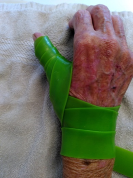
Medical Flossing bei Rhizarthrose Eine prospektive Studie mit 30 Patienten mit Follow up
Nicole Plüss, St. Gallen; Susanne Habelt, St. Gallen; Jörg Grünert, Goldach
DetailsHintergrund: Der Daumen und das Daumensattelgelenk sind von grosser Bedeutung und bei vielen Bewegungen im alltäglichen Leben unerlässlich. Bei Rhizarthrose kann es zu Schmerzen und Funktionsbeeinträchtigungen kommen. Als Therapieoption stehen eine ganze Reihe von konservativen und operativen Therapiemassnahmen zur Verfügung. Das Medical Flossing ist eine Therapie, die ursprünglich aus dem Spitzensport kommt und erst in den letzten Jahren in die Handtherapie Einzug gehalten hat. Das Medical Flossing regt Regenerations- und Reparaturmechanismen an. Fragestellung: Bei der Daumensattel-gelenksarthrose wird das Medical Flossing mit einer Wiederholung pro Sitzung empfohlen. Sind mehrere Wiederholungen effektiver und nachhaltiger bezüglich Schmerzlinderung, der Verbesserung des Bewegungsausmasses und der Qualität der Beweglichkeit? Methode: Es handelt sich um eine prospektive Studie, mit 30 Patienten,25 Frauen und 5 Männer, im Alter zwischen 37 und 61 Jahren. die innerhalb eines Jahres in die Studie aufgenommen wurden, unabhängig vom Schweregrad der Rhizarthrose. Im ersten Monat der Therapie erhielten die Patienten der Gruppe A zwei Wiederholungen und die Patienten der Gruppe B drei Wiederholungen des Medical Flossings pro Therapiesitzung. Im zweiten Monat erhielten dann beide Gruppen A und B jeweils drei Wiederholungen der Flossing Therapie pro Therapie Sitzung. Es wurden neben ROM und Kraft (Jamar, Pinch) auch ein Quick DASH score sowie eine Schmerzeinschätzung (VAS 1-10) vor und nach der Therapie und die Qualität der Beweglichkeit erfasst. Nach 6 - 12 Monaten erfolgte eine Nachkontrolle, darin konnten 28 Patienten erfasst werden. Ergebnisse: Hinsichtlich der Gelenksbeweglichkeit zeigte die Gruppe, welche drei Wiederholungen pro Sitzung hatte eine deutliche Verbesserung Bewegungsqualität, der Kraft und der Schmerzen. Der Quick DASH score sank von 54 und 25 am Ende der Therapie. Durchschnittliche Nachkontrollzeitpunkt war 8 Monate (6 - 12Monate) nach abgeschlossener Flossing Therapie. Es wurden 28 Patienten nachkontrolliert. Bei allen Patienten waren die Verbesserungen immer noch klar feststellbar. Schlussfolgerung: Diese Studie zeigt, dass eine repetitive Anwendung des Medical Flossing zu einem nachhaltigen Erfolg betreffend Bewegungsqualität und einer anhaltenden Schmerzreduktion führt. Alle 28 Patienten waren mit der konservativen Therapie des Medical Flossings sehr zufrieden und würden diese Therapiemethode wieder wählen.
|
|
|
|
FM74
Betätigungsbasierte Handtherapie – Tipps und Tricks für den Arbeitsalltag
Bernadette Tobler-Ammann, Bern; Astrid Schmid, Thun
DetailsDer Beruf der Ergotherapie hat die Beschäftigung als zentrales therapeutisches Therapiemittel übernommen und angewendet. Dieses Konzept wurde auch in die Physiotherapie und Handtherapie integriert. Eigentlich erscheinen betätigungsbasierte Interventionen sowohl logisch als auch praktisch, um diese in Behandlungsprogrammen in verschiedenen klinischen Settings einzubeziehen 1. In der Kultur des biomechanischen Modells im Gesundheitswesen ist es jedoch eine Herausforderung, betätigungsbasierte Interventionen in der Handtherapiepraxis zu integrieren. Unter einer betätigungsbasierten Intervention wird eine zweckgerichtete und bedeutungsvolle Aktivität für eine Person in ihrem sozialen Kontext verstanden 1. Zurzeit gibt es noch wenig Literatur betreffend Wirksamkeit und Integration von betätigungsbasierten Interventionen in der Behandlung von HandpatientInnen. Eine Übersichtsarbeit zum Thema zeigt auf, dass die vorhandene Literatur vielversprechende Trends im Einsatz von Betätigung als Behandlungsintervention für muskuloskelettale Beschwerden der oberen Extremität aufzeigt 2. Patienten gaben zum Beispiel an, zufriedener mit dem Behandlungsergebnis zu sein, da dieser kundenorientierte Ansatz hilft, die persönlichen Bedürfnisse des Patienten besser zu identifizieren. So werden die Wiedererlangung gewünschter Aktivitäten und Rollen zum Therapieziel, welches gemeinsam zwischen Therapeut und Patient entschieden und priorisiert wird. Zudem kann das Ausführen von Aktivitäten von der verletzten Hand ablenken und so deren Einsatz im Alltag verbessern. Wie können denn nun betätigungsbasierte Interventionen in der Handtherapie umgesetzt werden? Sind diese überhaupt sinnvoll? In diesem Beitrag möchten wir über Herausforderungen und Chancen dieses Therapieansatzes in der Handtherapie sprechen und Tipps und Tricks für dessen Einsatz im Arbeitsalltag aufzeigen, wie zum Beispiel dem Überdenken des eigenen Einsatzes von Assessments oder der Suche nach Gleichgesinnten zum Austausch. 1. Valdes K, Naughton N, Téllez RC, Szekeres M. The use of occupation-based interventions and assessments in hand therapy: A cross-sectional survey. J Hand Ther. Dec 28 2021;doi:10.1016/j.jht.2021.10.008 2. Weinstock-Zlotnick G, Mehta SP. A systematic review of the benefits of occupation-based intervention for patients with upper extremity musculoskeletal disorders. J Hand Ther. Apr - Jun 2019;32(2):141-152. doi:10.1016/j.jht.2018.04.001 |
|
|
|
15:15 – 16:30
|
Freie Mitteilungen IIIInnovation |
|
|
Club Casino
SGH-FM3
|
Vanessa Reischenböck, Zürich; Michaela Winkler, Lausanne
|
|
|
FM20
Corrective Cold Ablation Robot-Guided Laser Osteotomies in Wrist Surgery – a cadaver study
Maximilian Hofer, Liestal; Florian Thieringer, Basel; Enrico Coppo, Bruderholz; Marta Morawska, Basel; Magdalena Müller-Gerbl, Basel; Philipp Honigmann, Bruderholz
DetailsPurpose The purpose of this study is to show accuracy of a Cold Ablation Robot-Guided Laser Osteotome in pre-clinical cadaver tests performing osteotomies in the field of wrist- and forearm surgery.
Methods Osteotomies were performed with CARLO® which is a miniaturized ablation laser with an optical system controlled by a navigation system. The energy of a laser pulse hitting the bone tissue heats up the water content of the bone and vaporizes it. The debris is being expelled immediately, providing a clean-cut line with preservation of the bone microstructure. CARLO® enables new cutting patterns that are impossible to achieve with conventional methods. A total of 12 corrective laser-osteotomies were performed on the distal metaphyseal radius and ulna. The osteotomies were planned patient specific on a 3D CT-model prior to surgery using CARLO®’s planning software. On the radii six two plane adjustments were conducted lengthening the radius by 4mm and correcting radial inclination by 10° using a curved sine cut. On the ulnae one plane corrective osteotomies were performed shortening the ulna by 3mm using a sine cut as well. Pre-and postoperative CT-scans were taken to compare the virtual surgical planning with the post-operative results and to show accuracy of the cutting path.
Results Accuracy measurements of the actual cutting path compared to the planned cut showed a mean deviation of 2.11mm in the osteotomies of the radii and 1.66mm on the ulnae. The inaccuracy could have several possible causes, most probable is that the surface registration was not done careful enough. However, the resulting corrections showed great precision being at least as accurate as todays techniques using 3D printed patient specific cutting guides and 3D planning methods. After fixation the radii revealed an average lengthening of 4.36mm (planned: 4.00mm), while radial inclination was corrected by 10.55° (planned: 10.00°). The ulnae showed an average correction of 3.05mm (planned: 3.00mm).
Conclusion First cadaveric results show promising results concerning the accuracy of one and two plane osteotomies in wrist and forearm surgery using a Cold Ablation Robot-Guided Laser Osteotome. Accuracy testing showed that registration of the bone in relation to the patient marker is key to achieve a high precision, therefore it should be done carefully. Future steps are stability testing of the osteosynthesis followed by certification and first use in patients. |
|
|
|
FM21
Preliminary data of a new integrated digital platform for remote rehabilitation in hand surgery
Maurizio Calcagni, Zürich; J Rodriguez, Barcelona (ES); Ricardo Jauregui Telleira, Barcelona (ES); Inga Besmens, Zürich
DetailsRehabilitation is a central part of the treatment of hand and wrist conditions and it represents the most time-consuming part. Normally, rehabilitation take place partially in the office under the control of the therapist and partially alone at home. This second part is left to the compliance of the patient in term of number of sessions per week and of quality of the exercises. Remote rehabilitation can be a good solution to overcome these problems if a feed-back loop can be established between all the involved stakeholders. ReHub®, a new digital platform for remote rehabilitation that connects the surgeon, the therapist and the patient was developed to fulfil these needs. Based on the clinical information given by the physician, the therapist creates a personalised programme. The platform allows for an automatic tracking of the movements, the data recorded during the exercise are uploaded in the cloud and are used to monitor the improvements. The patient can answer to different questionnaires (PROMs). During the exercise the patient can interact with the platform to learn and monitor the right way to perform the exercises. In 2021-2022. 50 patients were treated with ReHub® for different upper limb pathologies. Patients received a programme of 3 sessions a week for 4 weeks. The number of sessions executed, the Quick-DASH and the pain levels were measured at the beginning and after 2 and 4 weeks. The diagnosis treated with ReHub® were 15 distal radius fracture, 10 carpal tunnel release and synovitis, 5 wrist sprain. The compliance rate was 66% and the drop-out rate was 6%. 34 patients filled the QuickDASH and its score improved in all cases of at least 15 points. Pain decreased steadily with an overall reduction of moderate (VAS 4-6) and intense (7-10) from 30.3% at the beginning, to 23.2% at 2 weeks and 6.9% after 4 weeks. The ReHub® digital platform demonstrated a very high compliance with a very low drop-out rate of only 6%. QuickDASH value improved and pain was reduced significantly. These findings reflect the high satisfaction rate of this new digital tool. Physicians can follow-up the improvements, not only with the automatic movement recognition tool, but also through the PROMs filled out by the patients. The therapists can evaluate the compliance in term of time spent exercising and the quality of the movements. The system proved effective in the rehabilitation of common upper extremity conditions, with a high compliance and patients' satisfaction. |
|
|
|
FM22
Biomechanical Stability and Viability of 3D Printed Bioresorbable Implants for Distal Radius Osteotomy
Alissa Gübeli, Genève; Adam Jakimiuk, Basel; Michaela Maintz, Basel; Florian Thieringer, Basel; Marco Keller, Zürich; Philipp Honigmann, Bruderholz
DetailsObjective: The fixation of distal radius osteotomy traditionally involves the use of titanium plates and screws, which has certain drawbacks such as soft tissue irritation, the need for removal in many cases, stress shielding, and potential patient intolerance or allergic reactions. In this study, we propose a new surgical technique utilizing 3D printed patient-specific implants made of bioresorbable material. We conducted an in vitro study to evaluate the biomechanical stability, biodegradability, and viability of these implants compared to standard titanium plates. Methods: We performed an opening wedge osteotomy of the distal radius on five cadavers. The osteotomy was planned virtually using CT scans of the radii and executed with the assistance of 3D printed cutting guides. Cadaver-specific 3D printed wedges made of poly(L-lactide-co-D, L-lactide) with 30% β-tricalcium phosphate (PLDLLA/β-TCP) were inserted into the osteotomy gap. They were designed with a hole for fixation using Arthrex Bio-compression screws (3-3.7 mm). 3D printing was performed using the Arburg Plastic Freeformer. Biomechanical tests were conducted to assess the strength of the fixation technique using the printed implants, comparing them to titanium plate implants. Mechanical tests involving tensile, compression, and bending forces were conducted on the printed implants to evaluate their mechanical performance. Biodegradation studies were carried out by submerging the implants in phosphate buffer solution (PBS) for up to 4 weeks. Results: Biomechanical investigations indicated that the fixation method utilizing the 3D printed implants could withstand an average axial force of 1879.7 N, which is lower than the titanium plate (2415.55 N) but a load higher than typically encountered during falls. They thus have the potential to provide sufficient stability as an alternative to standard titanium plates. However, physico-chemical tests revealed material alterations caused by the 3D printing process. Conclusion: Preliminary results of this study demonstrate promising biomechanical stability and viability of 3D printed bioresorbable implants for distal radius osteotomy. Since the 3D-printing process induced material alterations, further research is required to comprehensively evaluate the long-term viability and safety of this technique. If proven successful, these patient-specific implants could offer a beneficial alternative to traditional titanium plates. |
|
|
|
FM24
Long-term follow-up after digital nerve reconstruction with silk fibroin nerve conduit
Laima Bandzaite, Zürich; Olga Politikou, Zürich; Inga Besmens, Zürich; Martina Greminger, Zürich; Maurizio Calcagni, Zürich
DetailsIntroduction Multiple techniques are available for reconstruction of digital nerve defects using autograft, allograft and conduits. So far, comparative studies demonstrate variable results ranging from no difference in sensory outcomes to superior recovery with allograft and autograft reconstruction. SILKBridge®, a novel hybrid three-layered tube based on silk fibroin (obtained from the silk of Bombyx mori) was developed with optimized characteristics for peripheral nerve regeneration. The device has two electrospun layers (inner and outer) and an intermediate textile one. The first in-human pilot study of digital nerve reconstruction with SILKBridge® was performed in our institution and has proven its feasibility, safety and efficiency in the mid-term follow-up. We examined the long-term results in terms of function, patient satisfaction and biological behavior of the silk fibroin device. Patients and methods This is a follow-up study including patients with digital finger reconstruction with SILKBridge® following traumatic injury or neuroma resection. We collected data from last follow-up during routine clinical controls. Two-point discrimination, Tinel sign, local soft tissue conditions and pain were assessed. Ultrasound examination was performed for long term evaluation of conduit integration properties. Results In total four patients were included with median follow-up time of 3 years (1-3.5). All patients showed a good sensation recovery with a static 2-point discrimination of 5-12mm, a moving 2-PD of 4-10mm. No local signs of chronic inflammation or foreign body reaction were observed and a Tinel sign was absent in all patients. The scar was soft and the digital range of motion was the same on both sides. All patients were pain free and satisfied with the result, while no one reported a foreign body sensation. The silk conduit was still visible in the ultrasound but without signs of scar tethering or soft tissue reaction. Discussion This is the first long-term follow-up after digital nerve reconstruction with the hybrid, multi-layered silk fibroin device SILKBridge®. So far, functional results and patient satisfaction are excellent with no adverse events. SILKBridge® appears to be a safe and efficient alternative to nerve autografts or allografts in cases of digital nerve defects with very good long-term biological behavior. |
|
|
|
FM25
Ultrasound controlled mini-invasive A1 pulley release using a new guide instrument - a case series
Nora Schlimme, Bern; Esther Vögelin, Bern; Damian Sutter, Bern
DetailsObjectives: With percutaneous pulley release becoming more popular, safety and reliability of this mini-invasive procedure remain a concern. Visualization of the surgical steps by ultrasound suggests increased safety but also highlights the proximity of tendons, nerves and vessels as well as the potential for damage to these structures. Therefore, proper instrumentation is crucial. We present the results of implementing sonographically guided mini-invasive A1 pulley release using a newly designed guide instrument and a commercial hook knife.
Methods: Initially, basics for the technique using an early version of the guide instrument were established in cadaver hands. The instrument design was subsequently refined. Between November 2019 and December 2022 a total of 128 sonographically guided A1 pulley release procedures were performed in 79 patients. The guide instrument is inserted through a small incision at the base of the finger and enters the flexor tendon sheath between the A1 and A2 pulley. It is advanced proximally between the flexor tendon and the A1 pulley under sonographic visualization. Once in position, the hook knife is inserted through the channel in the guide instrument. The central position over the pulley is sonographically confirmed, before turning the hook knife 90°. Finally, the pulley is released by retracting the hook knife distally.
Results and Conclusions: Complications include one case of inadvertent skin laceration, one case of postoperative infection and one CRPS. Postoperative recurrence of a trigger finger due to incomplete pulley release was noted in 3 cases, in one of which the issue was resolved sonographically. The second had a remission after cortisone infiltration while the last underwent open pulley release revealing an intact A1 pulley. Intraoperative conversion to open release was performed in 5 cases due to an unfavorable position of the hook knife or persistent trigger finger intraoperatively. Of the 79 operated patients 68 (106 of 128 A1 pulley releases) reported return to strenuous activities within two weeks. No injuries to nerves, vessels or tendons occurred. In conclusion, the choice of appropriate surgical instruments and practice allows for safe and efficient implementation of a mini-invasive procedure in pulley release. |
|
|
|
FM26
Biodegradable Temporising Matrix - a reliable flap alternative for reconstruction extremity defects
Lena Fuest, Zürich; Esther Vögelin, Bern
DetailsIntroduction: Treatment of skin defects on the hand with exposed tendons, nerves or bones after severe trauma/infections can be a reconstructive challenge. Biodegradable Temporising Matrix (BTM®) may serve as a new soft tissue cover alternative if skin grafts alone do not heal and flaps are not an option, especially in patients that are not ideal candidates for major complex microsurgical reconstruction. BTM is a synthethic polyurethane dermal substitute. This case series represents the first study evaluating the efficacy of BTM in no-burns reconstruction. Method: In the year 2022, 27 patients were treated with BTM. In 20 cases the soft tissue defect was caused by a severe trauma. In four patients, there was a skin defect after an infection and in one case each, it was caused by an epitheloid sarcoma, by Dupuytren’s disease and by MDA5 Dermatomyositis. 59% of the patients were male, the median age was 51 years. Four patients suffered from an amputation of a finger, 18 patients had a substantial defect at the dorsum of the hand, three patients at the forearm, and one patient each had a defect at the thenar or palm. The size of the skin defect ranged from 1 to 200 cm2. Following debridement, BTM was applied to the wound. After the granulation tissue has fully integrated into the BTM layer, the sealing membrane is removed and a new, vascularized dermal layer emerges. A split-thickness skin graft was then applied. Results: On average, the delay between covering the soft tissue defect with BTM and the application of a split-thickness skin graft was 36 days. BTM was applied in 9 cases on exposed extensor tendons, in 4 cases on muscle or soft tissue, in 5 cases on bone, in 2 cases on the neurovascular bundle and in 1 case each on exposed flexor tendon and nerve transfer. One infection with enterobacter cloacae was observed. The split-thickness graft was well integrated after five days. At follow-up the ROM was satisfying according to the accompanying injuries. Conclusion: According to the statistics and experience gained, the use of BTM demonstrates enormous potential in healing complex wounds. It shows reliable healing and the scars after BTM application and split thickness skin grafts were aesthetically very pleasing. Coverage of tendons, nerves and bones are possible as well as coverage of infected wounds. The method should be preferred especially in patients, who do not qualify for a vascularized free or pedicled flap with reduced mobility requirements. |
|
|
|
FM27
Automated Detection and Localization of Distal Radius Fractures using a Convolutional Neural Network
Marco Keller, Zürich; Philipp Honigmann, Bruderholz; Cédric Huwyler, Windisch; Martin Melchior, Windisch; Florian Thieringer, Basel
DetailsIntroduction: Deep Learning and especially Convolutional Neural Networks (CNN) have established themselves as state-of-the-art methods in the field of image and object detection throughout the last decade. In health care they are successfully used, for example, to detect skin or breast cancer, where they reach the level of an expert opinion. The aim of this study was to build an deep learning algorithm which is able to automatically recognize the region of interest on standard radiographs, automatically identify and localize distal radius fractures. Material/Method: A database of conventional radiographs taken from 729 patients was manually built and labelled to serve as ground truth. 409 of these patients sustained a distal radius fracture (verified with X-rays, CT scans and clinical information from follow-up-appointments) and 320 were healthy individuals. By using all wrist X-rays from one institution from one calendar year we achieved a close-to-reality, wide variability of fracture configurations and patient demographics. The dataset was divided into a training- and a test-set. Several object detection deep learning models were trained to recognize the region of interest, to detect and localize distal radius fractures (if present). Furthermore, we explored if using one or two projections (dorsopalmar and lateral) and the addition of demographic data (age, gender) to the algorithm would change the models performance. The CNN model was compared to human observers in order to explore the clinical relevance. Results: Our best model detected distal radius fractures on dorsopalmar radiographs with an accuracy of 98.5% and an AUC of 0.995 improving on the human baseline accuracy by 18.5%. This corresponds to a sensitivity of 0.987 and a specificity of 0.987. The addition of a second projection (lateral) or demographic data didn’t improve the performance, but the model became more confident in its prediction. The region of interest was reliably recognized using automated detection of three landmark points which led to the inclusion of 99% of all fractures. Conclusion: Our deep learning model showed high accuracy in the detection and localization of distal radius fractures. In our test setup, it exceeded the performance of human observers. These kinds of Convolutional Neural Networks have the potential to improve the efficiency of healthcare workers and substantially change our way of patient care in the near future. |
|
|
|
FM28
A new grip strength measurement device – considering handle diameters and forearm rotations
Gabriella Fischer, Zürich; Maurizio Calcagni, Zürich; Tassilo Schirmer, Zürich; Pascal Schütz, Zürich
DetailsIntroduction: Methods:
Reliability of the HGS (ICC =0.96, range [.90, 99]) for all observed conditions as well as concurrent validity and device correlation (r=0.91 [.71, .99]) were found to be excellent. Between-session MDD were 83.6N, 85.5N and 32.5N for the 30mm, 60mm and 100mm HGS, respectively, which constitutes of 12.1-16.0% of measured force. Grip forces were found significantly different between handle diameters (p<0.0001) and forearm positions (p<0.0003). Across all positions, the smaller the diameter, the larger the observed gripping force. Interestingly, the grip force with HGS 30mm was larger in supination than in neutral position while using HGS 60mm, the highest grip force was reached in the neutral position, followed by supination. Measured grip strength significantly depends on the diameter of the handle and forearm rotation. As known from the ulnar impaction syndrome, pathologies can have an increased influence on grip strength in different positions. We therefore point out the necessity to investigate non-neutral positions and different handle diameters in order to understand healthy and pathological power grip force and its implication on daily activities. The presented HGS with its cylindrical design in relevant diameters for daily activities are mobile, easy to use, and now proved to be reliable and valid to be used in a clinical setting. They allow continuous recording of grip force and advanced analysis of grip force characteristics, such as loading-rate or centre-of-pressure, to get a more in-depth understanding of grip force characteristics in different pathologies. |
|
|
|
15:15 – 16:30
|
SGH Handfacts-Politpodium |
|
|
Theatersaal
SGH-HP
|
Max Winiger, Zürich
|
|
Politpodium Expertenrunde
Yvonne Gilli, Wil SG; Urs Hug, Luzern; Gian Signorell, Zürich; Walter Stüdeli, Wohlen bei Bern
Experten
|
||
|
16:30 – 17:00
|
Kaffeepause |
|
|
17:00 – 18:30
|
Mitgliederversammlung SGH |
|
|
Theatersaal
MV-SGH
|
||
|
17:00 – 18:30
|
Mitgliederversammlung SGHR |
|
|
Ballsaal
MV-SGHR
|